CT angiography a newer and less invasive technique is sometimes used. However the limbic system is still discussed in many traditional biology and physiology courses as part of the nervous system.
Nervous System Overview
The development of advanced modern technology has allowed researchers to build a more accurate understanding of how our brains work.
. The belief that the brain is the organ that controls behavior has ancient roots dating to early civilizations that connected loss of function to damage to parts of the brain and spinal cord. To study the nervous system a number of methods have evolved over time. We will begin with a general description of intracellular signaling events and why they are useful for understanding the properties of neurons.
Determine FROM WHERE a region receives its inputs determines the afferents by injecting tracer that selectively binds to surface GLYCOPROTEINS on axons and is take up by endocytosis and taken back to soma. Functional magnetic resonance imaging fMRI Nervous system. But these methods have some limitations.
Biochemical techniques are used to map out the various neurotransmitter systems in the nervous system. Up to 10 cash back Abstract. Brain scans include several types of imaging techniques used to diagnose tumors blood vessel malformations stroke injuries abnormal brain development and hemorrhage in the brain.
The static imaging technique is used to image the living brain. Brain slice preparations have most frequently been employed to answer questions of a biochemical or physiological nature. These methods include examining brain lesions microscopy electrophysiology electroencephalography and many scanning technologies.
The large electrical currents used during ECT create short seizures so patients who receive ECT must be monitored during treatment to ensure their safety. Types of brain scans include computed tomography CT magnetic resonance imaging MRI positron emission tomography PET and single proton emission SPECT scans. The nervous system uses a variety of neurotransmitter molecules but each neuron specializes in the synthesis and secretion of a single type of neurotransmitter.
Depression for example can be linked to levels of neurotransmitter and drug therapy for depression alters those levels. Next we will describe two fundamental methods used to study proteins. Transcranial magnetic stimulation TMS is a procedure in which magnetic pulses are applied to the brain of living persons with the goal of temporarily and safely deactivating a small brain region.
Nowadays due to advancements in technology scientists can use advanced imaging techniques to know more about the brain structure and the brain function. Imaging techniques are a newer and exciting way of studying the brain. This test takes a sample of cerebrospinal fluid from the spinal cord for testing.
Research using these techniques has found for instance that there are specific neurons known as feature detectors in the visual cortex that detect movement lines and edges and even faces Kanwisher 2000. Lesson Summary The limbic system is a set of structures in the brain. In TMS studies the research participant is first scanned in an fMRI machine to determine the exact location of the brain area to be tested.
Neuroscience is the scientific study of the nervous system the brain spinal cord and peripheral nervous system and its functions. Static Imaging and Functional Imaging. Producing and using antibodies and purifying proteins of interest.
These technological methods include the encephalogram EEG magnetic resonance imaging MRI functional magnetic resonance imaging fMRI and positron emission tomography PET. It is important to realize that these brain imaging techniques are not used for. Fifty years ago surgeons had to perform exploratory surgeries in order to diagnose brain.
Uses physiological techniques such as stimulation with electrodes to analyze and study various nervous system functions. In the brain neural activities often lead to metabolic activities such as increased blood flow and oxygen supply to the local vasculature. Electroconvulsive therapy ECT involves stimulating the brain with large amounts of electricity.
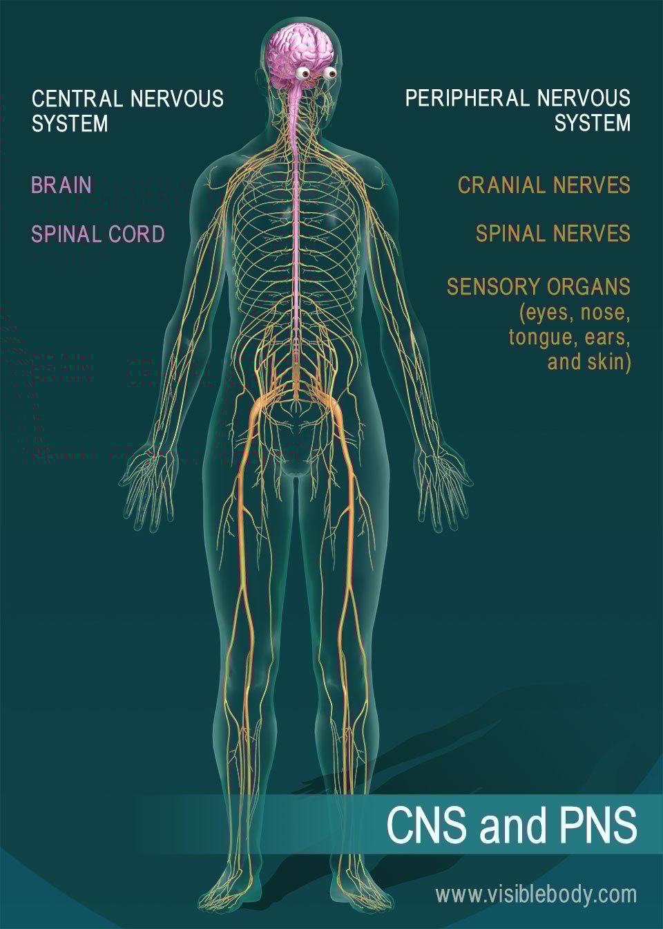
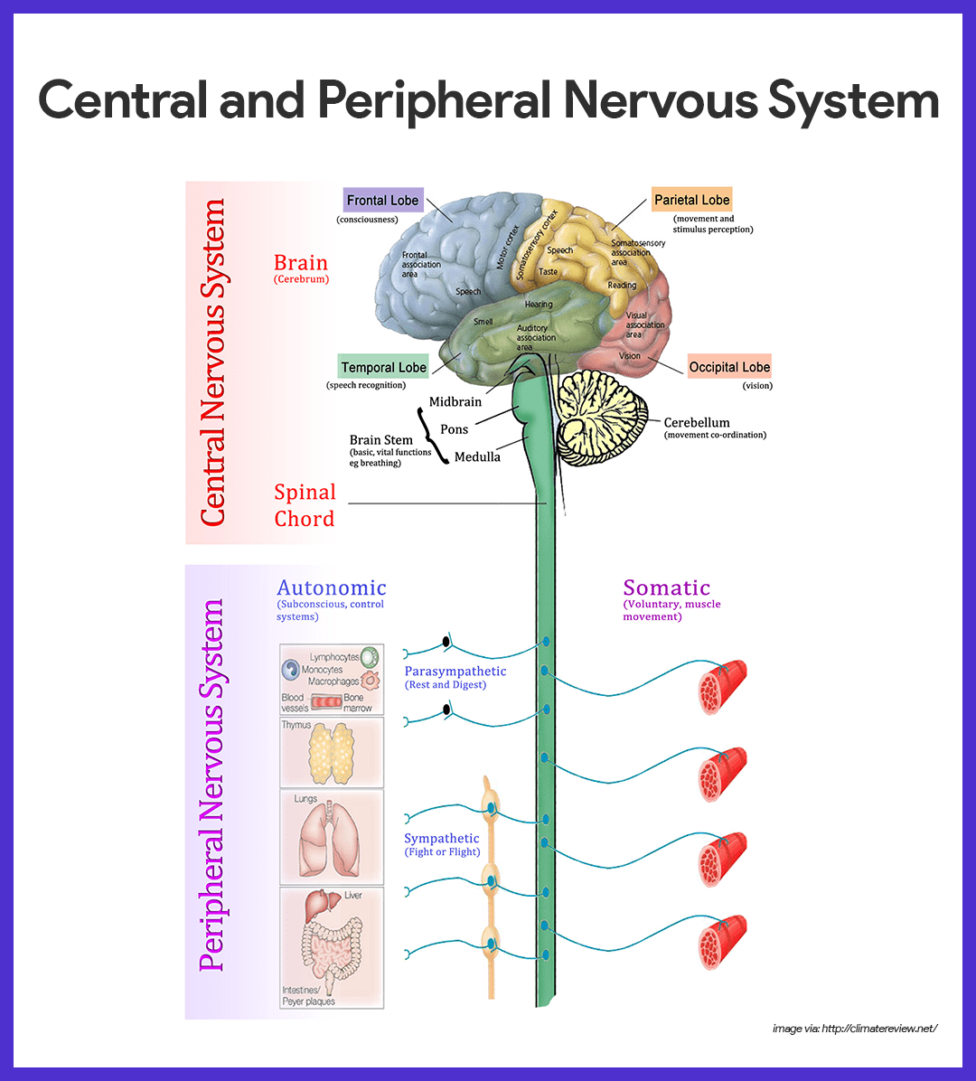
Name the various parts of the nervous system and their respective functions. Research using these techniques has found for instance that there are specific neurons known as feature detectors in the visual cortex that detect movement lines and edges and even faces Kanwisher 2000. Contrast fMRI requires injection of contrast.
Nevertheless in vitro brain slices also have considerable value as pharmacological tools with which to study the physiological actions of neurotransmitters and drugs on the central nervous system. It is one of the oldest techniques for treating problems in neural circuits. Several techniques can be used to detect changes of metabolic activities following neural activities including contrast fMRI blood-oxygen-level dependent BOLD fMRI and perfusion fMRI.
Immunohistochemical techniques IHC and in situ hybridization ISH are widely used techniques to study the expression of proteins and messenger RNAs in tissues and are extremely important to confirm and interpret biochemical and molecular results from the same tissues. But new imaging procedures enable scientists to study the brain in living animals including humans. These techniques ultimately have.
Paleoneurology employs analysis of prehistoric fossils to study the evoluation development and processes of the brain. There are two types of of imaging techniques. This test records the brains electrical response to visual auditory and sensory stimuli.
One approach primarily used with animals is to place detectors in the brain to study the responses of specific neurons. Explain how neurons communicate with each other. These experiments are usually combined with the neu-roimaging techniques well describe in a moment so that the investigators can match a precise picture of the persons brain damage with an equally precise assessment of her deficits.
Cerebral spinal fluid analysis also called spinal tap or lumbar puncture. One approach primarily used with animals is to place detectors in the brain to study the responses of specific neurons. Modern imaging methods such as MRI Magnetic Resonance Imaging scans use strong magnetic fields and radio waves to show details of brain structure and function.
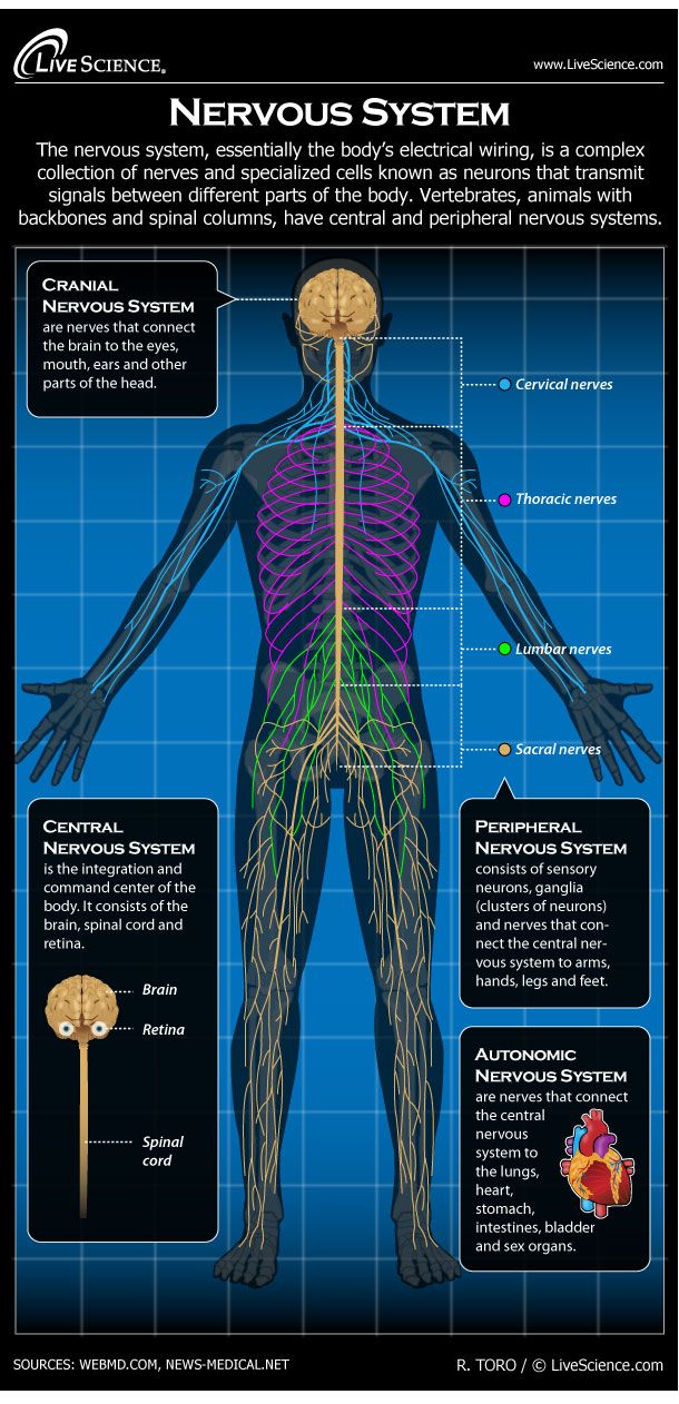
Human Nervous System Diagram How It Works Live Science

The Nervous System Special Senses
Nervous System Anatomy And Physiology Nurseslabs


0 Comments